- Home
- Others
- Others News
- 3D printed liver replica to help in transplant operations
3D-printed liver replica to help in transplant operations

Till date, surgeons look at a magnetic resonance image (MRI) or a computed tomography (CT) scan to visualise the liver and plan the operation.
"We provide the surgeons with a physical model that is 100 percent identical to what they would encounter in surgery when they operate," Nizar Zein, chief of hepatology at the Cleveland Clinic in Ohio, was quoted as saying.
It takes away some of the potential surprises that would be found at the time of surgery.
To create the artificial liver, the researchers combined the MRI and CT scans that patients have already undergone and then recreated the 3D shape of the organ.
These models were anatomically accurate in terms of volume and location of vessels in the liver.
Using these models, the team created the 3D-printed organs using a transparent polymer, then dyed the main blood vessels and the bile ducts.
The researchers are now developing similar methods to guide complicated surgeries, such as hand and face transplants, and pancreatic tumour removals.
The new liver replica could also be used to train medical students in the techniques needed for surgery, Zein added in a study published in the journal Liver Transplantation.
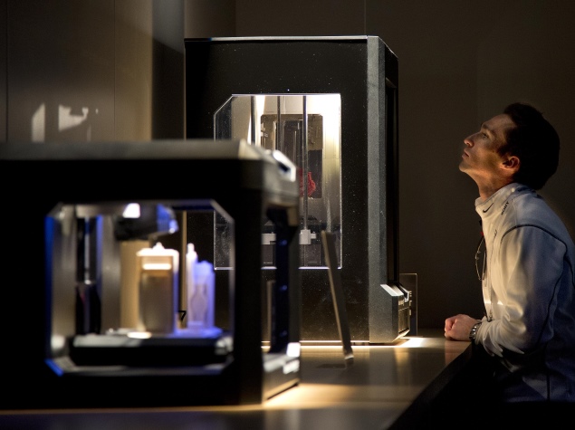
Relevantly, a US company in January claimed to have developed the world's first multi-material full-colour 3D printer capable of making objects of hard, soft and flexible polymers.
The 3D printer developed by Stratasys features "triple-jetting" technology that combines droplets of three base materials, reducing the need for separate print runs and painting.
Written with inputs from IANS
Catch the latest from the Consumer Electronics Show on Gadgets 360, at our CES 2026 hub.
Related Stories
- Samsung Galaxy Unpacked 2025
- ChatGPT
- Redmi Note 14 Pro+
- iPhone 16
- Apple Vision Pro
- Oneplus 12
- OnePlus Nord CE 3 Lite 5G
- iPhone 13
- Xiaomi 14 Pro
- Oppo Find N3
- Tecno Spark Go (2023)
- Realme V30
- Best Phones Under 25000
- Samsung Galaxy S24 Series
- Cryptocurrency
- iQoo 12
- Samsung Galaxy S24 Ultra
- Giottus
- Samsung Galaxy Z Flip 5
- Apple 'Scary Fast'
- Housefull 5
- GoPro Hero 12 Black Review
- Invincible Season 2
- JioGlass
- HD Ready TV
- Laptop Under 50000
- Smartwatch Under 10000
- Latest Mobile Phones
- Compare Phones
- OPPO A6c
- Samsung Galaxy A07 5G
- Vivo Y500i
- OnePlus Turbo 6V
- OnePlus Turbo 6
- Itel Zeno 20 Max
- OPPO Reno 15 Pro Mini 5G
- Poco M8 Pro 5G
- Lenovo Yoga Slim 7x (2025)
- Lenovo Yoga Slim 7a
- Realme Pad 3
- OPPO Pad Air 5
- Garmin Quatix 8 Pro
- NoiseFit Pro 6R
- Haier H5E Series
- Acerpure Nitro Z Series 100-inch QLED TV
- Asus ROG Ally
- Nintendo Switch Lite
- Haier 1.6 Ton 5 Star Inverter Split AC (HSU19G-MZAID5BN-INV)
- Haier 1.6 Ton 5 Star Inverter Split AC (HSU19G-MZAIM5BN-INV)


